Influence of the MgO-TiO2 Co-Additive Content on the Phase Formation, Microstructure and Fracture Toughness of MgO-TiO2-Reinforced Dental Porcelain Nanocomposites
Article information
Abstract
The influence of the co-additive concentration (0 - 45 wt% with an interval of 5 wt%) of MgO-TiO2 on the phase formation, microstructure and fracture toughness of MgO-TiO2-reinforced dental porcelain nanocomposites derived from a one-step sintering technique were examined using a combination of X-ray diffraction, scanning electron microscopy and Vickers indentation. It was found that MgO-TiO2-reinforced dental porcelain nanocomposites exhibited significantly higher fracture toughness values than those observed in single-additive (MgO or TiO2)-reinforced dental porcelain composites at any given sintering temperature. The amount of MgO-TiO2 as a co-additive was found to be one of the key factors controlling the phase formation, microstructure and fracture toughness of these nanocomposites. It is likely that 30 wt% of MgO-TiO2 as a co-additive is the optimal amount for MgTi2O5 and Mg2SiO4 crystalline phase formation to obtain the maximum relative density (96.80%) and fracture toughness (2.60 ± 0.07 MPa·m1/2) at a sintering temperature of 1000°C.
1. Introduction
Owing to their esthetics and excellent biocompatibility, porcelain-based ceramics containing a mixture of dispersed crystalline and glassy matrix phases have been widely investigated as restorative materials in dental applications. However, similar to other glass ceramics, the strong atomic bonds of their Si-O networks allow for no free electrons, resulting in brittleness of the material accompanied with low fracture toughness values in the approximate range of 1.01 – 1.40 MPa·m1/2.1,2) These values are too low for such materials to be used in dental applications, especially when extremely high strength and toughness are required.2) For example, three-unit fixed dental prostheses (FDPs) in the anterior region, posterior crowns, or core materials for crowns require fracture toughness values in the range of 2.25 – 2.75 MPa·m1/2.3,4) An effective method proposed to overcome these barriers involves the use of high-strength crystalline phases such as titania (TiO2), magnesia (MgO) or zirconia (ZrO2)1,5–7) in the form of a dispersion within the ceramic matrix via the formation of a composite structure. This finding was supported by Montoya et al.,8) who observed improved toughening properties in alumina porcelain ceramics after adding 2 wt% of TiO2. They suggested that the creation of mullite phases induced by TiO2 as an additive together with their dispersion within a glassy matrix acts as an important contributor to the strengthening of alumina porcelain ceramics. Tan et al.9) investigated the role of MgO as an additive on the fracture toughness of hydroxyapatite ceramics and found that the obtained fracture toughness value increased from ~ 1.48 ± 0.17 MPa·m1/2 to 1.64 ± 0.28 MPa·m1/2 after adding MgO from 1 wt% to 5 wt%, respectively. They explained that this observation could be attributed to the reduction of the grain size of the hydroxyapatite ceramics induced by the increased amount of added MgO. Apart from the type of additives, a potential improvement in the fracture toughness could be achieved for a number of dental ceramics by employing different amounts of oxide additives. However, to obtain greater fracture toughness, it is necessary to determine the optimal content for each material system. This finding was supported by Ragurajan et al.,10) who reported the effect of the MgO content (0.1 – 1 wt%) on both the density and fracture toughness of yttria (Y2O3)-stabilized tetragonal zirconia polycrystal ceramics. They found that when 0.3 wt% of MgO is added, ceramics reach their maximum density level, whereas after an addition of 1.0 wt% MgO, maximum fracture toughness is observed. They suggested that the increased fracture toughness resulted from the good stability of the tetragonal ZrO2 phase when the MgO content is increased from 0.5 to 1.0 wt%. Manshor et al.11) also investigated the effect of the TiO2 content (0 – 10 wt%) on the mechanical properties of zirconia-toughened alumina (Al2O3) composites. They found that the fracture toughness values increased significantly from 5.93 MPa·m1/2 to 6.56 MPa·m1/2 when the TiO2 content was increased from 0 to 3 wt%. The presence of TiO2 led to the development of elongated Al2TiO5 grains, which by some means greatly influenced the fracture toughness of the materials. These elongated grains cause a crack to travel along more than one plane and then to proceed around the grain, toughening the crack deflection via an intergranular fracture mode. Thus, this mechanism was suggested to be responsible for the improved fracture toughness.11) On the other hand, only minor degrees of improvement from 6.61 MPa·m1/2 to 6.64 MPa·m1/2 were observed with further TiO2 additions from 5 to 10 wt%. Based on these outcomes, both the type and content of oxide additives are likely to be key factors influencing the fracture toughness of these dental ceramics.
However, amongst all the approaches reported thus far, most attention has been concentrated on the use of a single additive, whereas investigations of the “co-additive” technique12) have not been widely reported. Furthermore, there is no report to date about the influence of the content of MgO-TiO2 as a co-additive on the structural and mechanical properties of MgO-TiO2-reinforced dental porcelain nanocomposites. Moreover, according to the phase diagram of the MgO-TiO2 system reported by Shindo,13) there are three possible stable magnesium titanate phases, Mg2TiO4, MgTiO3 and MgTi2O5 which possess high thermal stability and strength properties.14) Thus, it is believed that the formation of magnesium titanate phase(s) may result in toughening mechanisms suitable for obtaining improved fracture toughness of these dental porcelain composites. Moreover, TiO2 is typically used as a sintering aid added to promote low-temperature densification of MgO ceramics. Certainly, greater product density can be obtained along with lower overall processing costs.15) Therefore, this study aimed to investigate the influence of MgO-TiO2 as a co-additive on the phase formation, microstructure and fracture toughness of MgO-TiO2-reinforced dental porcelain nanocomposites.
2. Experimental Procedure
The materials used in this study were fabricated using a one-step sintering technique. Various amounts (0 - 45 wt% with an interval of 5 wt%) of MgO (lab grade), TiO2 (lab grade) (No. 5480MR, Nanostructured & Amorphous Materials, Inc., Houston, TX, USA) and MgO-TiO2 co-additives at a ratio of 1: 1 by weight were mixed with dental porcelain powders (Vita-VMK95, Vita Zahnfabrik, Bad Säckingen, Germany). In brief, green samples were obtained by mixing powders with polyvinyl alcohol (PVA) binder via a slip-casting technique, as suggested by the manufacturer.16) These were then poured into a standard stainless steel mold with a regularly sized cavity of 30 mm × 6 mm × 2 mm, reproducing the desired dimensions and shapes.1) After molding, the samples in the control group (0 wt% additive) was sintered at 980°C for five minutes at a heating rate of 25°C/min, as recommended by the manufacturer.16) In the other groups (5 – 45 wt% additive), the sintering temperature was designed based on a differential thermal analysis of the MgO-TiO2 mixture where an exothermic peak was found at ~ 800°C. Therefore, the composite samples were sintered at 800°C/60 min at a heating rate of 25°C/min16) in a vacuum furnace (Multimat Touch & Press, Hanau, Germany). The sintering temperatures were then varied to 1000 and 1200°C in order to determine the optimal firing condition for each group (MgO, TiO2 or MgO-TiO2 co-additives). The fracture toughness of all of the composites was determined using the Vickers indentation method as proposed by Anstis et al.17) Loads of 9.8 N from a Vickers microhardness tester (STARTECH SMV-1000, Guiyang Sunpoc International Trade Co., Ltd., Guiyang, China) were applied to thirty specimens per group. For each specimen, ten indentations were examined under a microscope, and the lengths of the cracks at the four corners of the pyramid were measured in order to calculate the fracture toughness (KIC) using the indentation strength method.
The equation proposed by Anstis et al.17) is as follows:
Here, E is Young’s modulus in GPa, Hv is the Vickers hardness in GPa, P is the indentation load (N), and c is the crack length (m). After the specimens underwent applied loads of 9.8 N from a Vickers microhardness tester, they were etched by applying 8 vol% of hydrofluoric acid for three seconds. The relationship between the crack propagation and microstructure with different amounts of additives was investigated by scanning electron microscopy (SEM) (JSM-840A 6335 F, JEOL, Tokyo, Japan) and energy dispersive X-ray spectroscopy (EDS) for a chemical composition analysis.
The theoretical density of the samples was calculated according to the rule of mixtures, and the final density after sintering was measured by the immersion method in deionized water using the Archimedes’ principle (ten specimens per group). The grain size and morphology of the crystalline phase in the sintered samples were determined by means of SEM. Thirty polished specimens for each group were selected randomly and sectioned with a carborundum disc into specimens 3 mm × 3 mm × 2 mm in size. The specimens were etched with 8 vol% of hydrofluoric acid for three minutes and coated with gold (20 nm) to observe the microstructure of the composites. To examine the phase formation of the MgO-TiO2-reinforced dental porcelain nanocomposites, the samples (two specimens per group) were ground into powder before a room-temperature X-ray diffraction (XRD) analysis (X’pert MPD, Philips Corp., Almelo, Netherlands) was conducted using Ni-filtered Cu Kα radiation of 0.154056 nm λ at 40 kV and 45 mA.
3. Results and Discussion
In order to compare the improvement of the fracture toughness of the dental porcelain composites according to the MgO-TiO2 co-additive dispersion in the porcelain matrix and that due to MgO and TiO2 single-additive dispersions, the fracture toughness values of the composite materials after adding 0 – 45 wt% of MgO, TiO2 and MgO-TiO2, sintered under various sintering temperatures were investigated. These results are presented in Fig. 1 and Table 1. The fracture toughness of the dental porcelain (0 wt% additive) was ~ 1.27 ± 0.03 MPa·m1/2, close to the value reported earlier by Pisitanusorn et al.1) In this study, the fracture toughness values of all of the composites, except for the MgO additive at 800°C, were significantly higher than those of the dental porcelain. This finding was attributed to the addition of metal oxide (MgO, TiO2 and MgO-TiO2 co-additives), which produced a high degree of the crystalline phase which increased the crack resistance. This finding is consistent with those reported in other studies.1,5–7) For MgO, at a sintering temperature of 800°C, the fracture toughness increased when the content of the additive was increased from 0 wt% to 20 wt%. A similar observation was made by Tan et al.,9) who found that the obtained fracture toughness value of hydroxyapatite ceramics increased after MgO was added. On the other hand, in this study, when the additive content exceeded 20 wt%, the fracture toughness values not only decreased sharply but the fracture toughness values in the groups with 40 and 45 wt% of MgO were also lower than those of the pure dental porcelain. This may have resulted from poor densification owing to the resulting nonhomogeneous ceramics when the additive concentration was too high.18) However, the fracture toughness values of MgO composites tended to increase when the sintering temperature was increased to 1000°C and 1200°C. This finding was consistent with the findings of She et al.,19) in their study of Al2O3- and Y2O3-doped SiC ceramics. They suggested that the increased fracture toughness may be due to enhanced crack propagation resistance stemming from the coarsening of the grains at a relatively high sintering temperature. In this study, the maximum fracture toughness (~ 1.87 ± 0.07 MPa·m1/2) of the MgO-reinforced dental porcelain composites was observed after adding 30 wt% of the MgO additive and sintering at 1200°C.
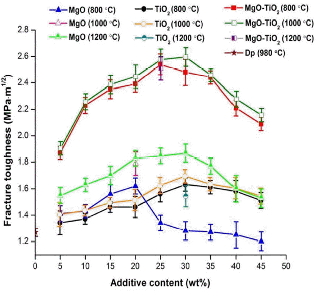
Fracture toughness of various additive contents of MgO, TiO2 and MgO-TiO2 as co-additives to reinforce dental porcelain composites.

Physical Properties of Dental Porcelain Nanocomposites Containing Various Amounts of MgO-TiO2 Co-additive
For the TiO2 additive with the 800°C sintering scheme, the fracture toughness continuously increased when 5 - 30 wt% of TiO2 additives were added, in accordance with the findings of Manshor et al.,11) who observed improved fracture toughness values in ZrO2/Al2O3 composites after adding TiO2. However, in this study, similar to MgO, the fracture toughness decreased after adding more than 30 wt% of TiO2 additive. This may have resulted from the nonhomogeneity and poor densification of the sintered ceramics, as described previously.18) When the sintering temperature was increased to 1000°C, the fracture toughness value of the TiO2 composite materials tended to increase, a finding consistent with that by She et al.19) The maximum fracture toughness of TiO2 single-additive dental porcelain composites (~ 1.69 ± 0.05 MPa·m1/2) sintered at 1000°C was obtained by adding 30 wt% of TiO2 additive. However, when the sintering temperature was increased to 1200°C, the fracture toughness value of the composite materials tended to decrease compared to those sintered at 800°C and 1000°C. This finding resulted from excessively high sintering temperature, resulting in an exaggerated grain size, poor densification, and high porosity, all of which control the mechanical properties of ceramics, such as the fracture toughness of composite materials, as reported clearly in the literature.20–23)
As detailed in Fig. 1, the maximum fracture toughness of the MgO-reinforced dental porcelain composites was greater than the toughness when TiO2 was used. However, under optimal conditions, the maximum fracture toughness values of both the MgO and TiO2 systems were still lower than the required fracture toughness value (range ~ 2.25 – 2.75 MPa·m1/2) for dental applications, especially when extremely high strength and toughness are required.2–4) Interestingly, when the MgO-TiO2 co-additive was employed to toughen dental porcelain ceramics, it was found that MgO-TiO2-reinforced dental porcelain nanocomposites exhibited significantly higher fracture toughness values than those observed in single-additive (MgO or TiO2)-reinforced dental porcelain composites at any given sintering temperature. The greater potential co-additive effect over that found in single-additive reinforced dental porcelain composites was in agreement with the findings of other studies.24,25) Furthermore, the fracture toughness of the MgO-TiO2-reinforced dental porcelain nano-composite samples generally increased, reaching a maximum value when sintered at 1000°C, similar to the findings in the TiO2 additive group. This finding was attributed to TiO2, which is typically used as a sintering aid to promote the low-temperature densification of MgO ceramics.15) This combination of MgO and TiO2 may permit a reduction of the optimal sintering temperature from 1200°C (MgO single-additive) to 1000°C, to obtain high fracture toughness in MgO-TiO2-reinforced dental porcelain nanocomposites. Thus, for a clear understanding of the variation of the fracture toughness values in MgO-TiO2-reinforced dental porcelain nanocomposites, this study concentrated only on the influence of the MgO-TiO2 co-additive content at only the sintering temperature of 1000°C.
As demonstrated in Fig. 1 and Table 1, MgO-TiO2-reinforced dental porcelain nanocomposites showed three regions of fracture toughness changes: rapid growth, slow growth and a gradual decrease. After adding 5 – 15 wt% of the MgO-TiO2 co-additive to the dental porcelain, the fracture toughness rapidly increased to 2.39 ± 0.07 MPa·m1/2 (rapid growth region). The fracture toughness tended to increase continuously until the maximum value of approximately 2.60 ± 0.07 MPa·m1/2 was reached after adding 20 – 30 wt% of the MgO-TiO2 co-additive (slow growth region). A similar observation was also made with regard to the change of the relative density versus the amount of MgO-TiO2 as a co-additive, where the relative density increased and reached a maximum (~ 96.80%) after 30 wt% of MgO-TiO2 co-additive was added (Table 1). This finding can be explained by the correlation between the enhanced densification (lower porosity) and the MgO-TiO2 co-additive content. It was proved to be difficult to obtain highly dense samples with the MgO-TiO2 co-additive at < 30 wt% because the samples could not be densified using the employed sintering conditions, as evidenced by the predominant porosity in the SEM images of the dental porcelain nanocomposites with 5 wt% MgO-TiO2 as a co-additive (Fig. 2(a)) over the composites with 30 wt% of the co-additive (Fig. 2(b)).16) However, after adding 35 - 45 wt% of MgO-TiO2 as a co-additive, the fracture toughness and relative density were found gradually to decrease to ~ 2.16 ± 0.05 MPa·m1/2 and 75.76%, respectively (the gradual decrease region). A similar observation was also made by She et al.19) regarding the effect of Al2O3/Y2O3 as an additive on the fracture toughness and relative density of SiC ceramics. From this study, it can be assumed that the increased MgO-TiO2 co-additive content resulted in an increase in the relative density, which thus increased the fracture toughness, in accordance with the findings of other studies.8,26–29) However, an excessively high MgO-TiO2 co-additive content has been found to result in a nonhomogeneous ceramic, leading to poor densification and resulted in a decreased relative density and increased porosity (Fig. 2(c)), followed by the decreased fracture toughness.18) Apart from relative density, other factors, such as the grain size and the formation of a new crystalline phase in the composites, can affect the fracture toughness of materials, as reported in previous studies.8,26–29) Consequently, SEM and XRD techniques were employed to measure the grain size and to identify the crystalline phases of these dental porcelain composites for a better description of the variation of the fracture toughness values.

Representative SEM micrographs showing the pore distribution of dental porcelain ceramics nanocomposites with various additive contents: (a) 5 wt% of MgO-TiO2 co-additive, (b) 30 wt% of MgO-TiO2 co-additive and (c) 45 wt% of MgO-TiO2 co-additive.
In a SEM analysis, the microstructural evolution of all MgO-TiO2-reinforced dental porcelain nanocomposite samples was examined, as shown in Fig. 3 (G1–9). The smooth surface of typical porcelain-glass ceramics, dispersed with elongated nano-reinforcements embedded in a nearly round micronic grain, termed “micro/nano composites,” as described in the ceramic-nanocomposite models of Palmero,30) was revealed. The properties of the “micro/nano composites” make them resistant to high temperatures and interruptions of crack motions through grains.31,32) As shown in Fig. 3 and Table 1, the grain sizes of the composites vary between 0.28 μm and 0.80 μm and are related to the content of the MgO-TiO2 co-additive. It can be observed that with an increase in the MgO-TiO2 co-additive content, larger grains are produced, in accordance with the findings of Santos et al.33) Each sample group consisted of mixed fine and coarse grain sizes. These observations can be attributed to the influence of the TiO2 additive, which acted as a grain growth inhibitor and binder, leading to variation of the grain size. The function of the grain growth inhibitor resulted in a fine grain size, whereas the function of the grain growth binder enlarged the grain size due to the binding force at the grain boundary.34) The group with 45 wt% of MgO-TiO2 as a co-additive had a maximum grain size range of 0.50 – 0.80 μm. The described microstructure of the MgO-TiO2-reinforced dental porcelain nanocomposites made crack propagation more difficult than in the microstructure of non-reinforced dental porcelain due to the fact that the energy required for crack formation was higher.1) Based on an analysis of the surface damage caused by the Vickers indentation on their surfaces, the 30 wt% MgO-TiO2-reinforced dental porcelain nanocomposites achieved greater fracture toughness than did the non-reinforced dental porcelain, as shown in the micrographs of the cracks in Fig. 4. The optical microscopic image of the crack formation in the pure dental porcelain (Fig. 4(a)) shows long, straight lines with the cracks emanating from the edges of the pyramid and emerging on the surface near the corner of the pyramid, as shown by the arrow. In contrast, the cracks in the 30 wt% MgO-TiO2-reinforced dental porcelain nanocomposites processed at 1000°C show meandering paths with bend as a result of crack deflection, as denoted by the arrows in Fig. 4(b).
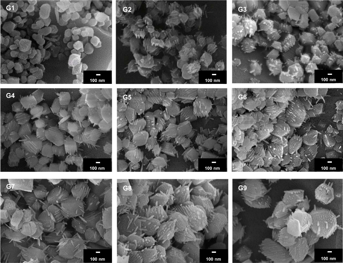
SEM of dental porcelain ceramic nanocomposites. The grain size increased with an increase in MgO-TiO2 co-additive content from G1 (5 wt%) to G9 (45 wt%), with an interval of 5 wt%.
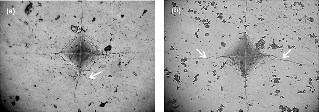
Representative micrographs of Vickers indentations: (a) crack propagation emanating from the lateral edge of the indent (arrow) in pure dental porcelain and (b) crack deflection (arrows) in 30 wt% MgO-TiO2-reinforced dental porcelain composites.
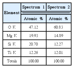
The microscopic dimensions of the crack after the sample with 30 wt% of MgO-TiO2 as a co-additive were etched (8 vol% of hydrofluoric acid, three seconds) in Fig. 5(a) shows that the crack path is clearly confined to the glassy matrix. In other words, the crack does not propagate through any of the crystals, as the crystals deflect the crack. To determine the chemical composition of these crystals, an EDS analysis of the 30 wt% MgO-TiO2-reinforced dental porcelain nanocomposites was conducted, as shown in Fig. 5(b), Fig. 5(c) and in Table 2. The EDS spectra were obtained from the crack-deflected crystals in spectrum 1 and spectrum 2, confirming the existence of O, Mg, Si and Ti. All key elements may be related to the composition of the MgO-TiO2 co-additive, and the presence of Si and O indicated the composition of a glass matrix.
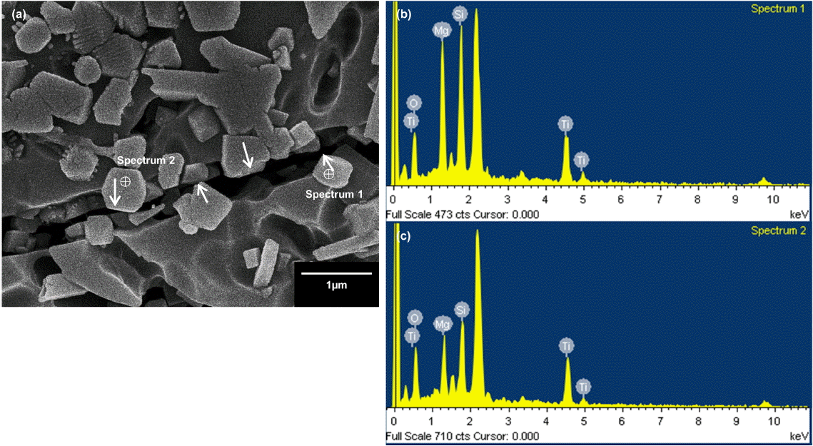
(a) SEM of crack deflection (arrows) of 30 wt% MgO-TiO2-reinforced dental porcelain ceramic nanocomposites, and (b) and (c) EDS analyses of crack-deflected crystals, indicated as spectrum 1 and spectrum 2 in (a), respectively.
Therefore, the increase in the crack surface area and the interlocking microstructure of the composites explain the high toughness of the composites. Thus, in this study, the crack deflection mechanism was primarily responsible for the effective toughening mechanism of the MgO-TiO2-reinforced dental porcelain nanocomposites.35) However, the literature has reported that the crack-deflection mechanism leads to only a 20 – 30% increase in the toughness, at most.19) In addition, the EDS technique can determine only the element composition in the SEM image. Therefore, the XRD technique was employed to analyze the formation of a crystalline phase, which may be another factor responsible for this enhanced fracture toughness of MgO-TiO2-reinforced dental porcelain nanocomposites.
The XRD patterns of the dental porcelain nanocomposites reinforced with different amounts of MgO-TiO2 as co-additives are displayed in Fig. 6. In the 5 wt% MgO-TiO2 co-additive group, the results identify the phases of MgO (JCPDS file No. 87-0653) and TiO2 (JCPDS file No. 73-1232), which are the starting precursors of the additives. Because the content of the MgO-TiO2 co-additive was excessively low, no trace of the formation of a new crystalline phase was detected in the 5 wt% MgO-TiO2 co-additive group. When the content of MgO-TiO2 as a co-additive was increased to 10 wt%, apart from MgO and TiO2, weak reflections of magnesium dititanate (MgTi2O5) peaks were detected. These could be matched with JCPDS file No. 79-0832, in agreement with the findings of other studies.36–38) The presence of MgTi2O5 is consistent with the phase diagram of the MgO-TiO2 system reported by Shindo.13) MgTi2O5 has well-balanced properties, such as low thermal expansion, high-temperature stability and potentially good mechanical properties. 39) Upon the first approximation, the fracture toughness in the group with 10 wt% of MgO-TiO2 as a co-additive increased significantly when compared to the group with 5 wt% of MgO-TiO2 as a co-additive, possibly resulting from the formation of the MgTi2O5 crystalline phase. When the MgO-TiO2 co-additive content was increased to 15 – 45 wt%, not only were peaks detected for MgO, TiO2 and MgTi2O5 phases, but new additional forsterite (Mg2SiO4) peak reflections were also detected, which could be matched with JCPDS file No. 34-0189, in agreement with the findings of other studies.40,41) In this study, Mg2SiO4 may have resulted from the combination of SiO2 in the glassy matrix of the dental porcelain2) and the MgO additive, a possibility consistent with the finding of Cheng et al.,42) who reported that Mg2SiO4 nanocomposites could be prepared from MgO and SiO2 mixtures. Additionally, Ni et al.43) reported that Mg2-SiO4 ceramics showed significantly improved fracture toughness when compared with hydroxyapatite ceramics, a finding which is consistent with the findings here. Thus, the EDS (Fig. 5(b), Fig. 5(c)) and XRD analyses (Fig. 6) confirmed the chemical compositions and the phase formation of MgTi2O5 and Mg2SiO4, respectively. In the 15 – 30 wt% groups, the fracture toughness increased until a maximum value of ~ 2.60 ± 0.07 MPa·m1/2 was reached, possibly resulting from the existence of both MgTi2O5 and Mg2SiO4 crystalline phases in the dental porcelain nanocomposites. On the other hand, although the 35 – 45 wt% groups consisted of both MgTi2O5 and Mg2SiO4 crystalline phases, the fracture toughness of the MgO-TiO2-reinforced dental porcelain nanocomposites was found to decrease to ~ 2.16 ± 0.05 MPa·m1/2. This reduction may have been due to the decreased relative density and increased grain size. Crack propagation in the 45 wt% MgO-TiO2-reinforced dental porcelain nanocomposites takes place not only within the glassy matrix but also through the crystals (arrows in Fig. 7). In other words, the larger grains are not strong enough to deflect incoming cracks. Therefore, crack propagation was described as transgranular crack propagation.
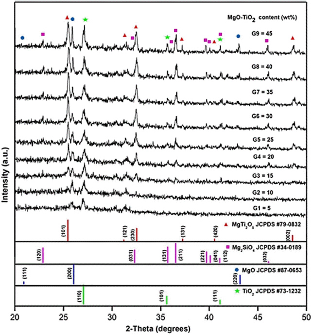
XRD patterns of dental porcelain reinforced with various amounts of MgO-TiO2 as a co-additive sintered at T = 1000°C.
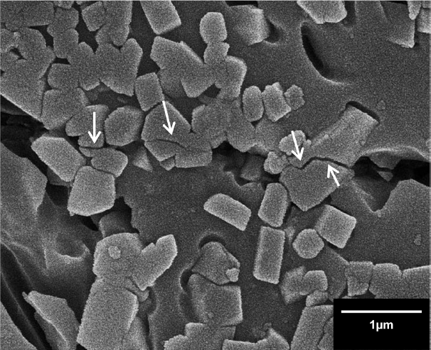
SEM of a transgranular fracture (arrows) in a 45 wt% MgO-TiO2-reinforced dental porcelain nanocomposites.
From our results, it can be concluded that the formation of multiple phases, including MgTi2O5 and Mg2SiO4 crystalline phases, may bring toughening mechanisms suitable for obtaining increased fracture toughness of MgO-TiO2-reinforced dental porcelain nanocomposites. In addition, the grain size and relative density also affected the fracture toughness of the composites. In this study, at an optimal sintering temperature (1000°C), 10 – 40 wt% of MgO-TiO2 as a co-additive provided fracture toughness values suitable for dental applications, in which high strength and toughness are required (~ 2.25–2.75 MPa·m1/2).2–4) Moreover, 30 wt% of MgO-TiO2 as a co-additive, producing MgTi2O5 and Mg2SiO4 crystalline phases together with the highest relative density among all groups, resulted in the maximum fracture toughness. Therefore, further studies should explore the effects of the content of MgO-TiO2 as a co-additive on other mechanical properties, such as the flexural strength and compressive strength, to enhance or tailor the key mechanical properties of dental porcelain-based materials.
4. Conclusions
The influence of the MgO-TiO2 co-additive content on the phase formation, microstructure and fracture toughness of MgO-TiO2-reinforced dental porcelain nanocomposites was investigated. The amount of MgO-TiO2 as a co-additive is a key factor when attempting to control the phase formation and microstructural evolution, considering that these factors affect the fracture toughness of MgO-TiO2-reinforced dental porcelain nanocomposites. The optimal content of MgO-TiO2 as a co-additive is 30 wt%, with sintering at 1000°C. This results in the formation of the MgTi2O5 and Mg2-SiO4 crystalline phases to obtain the maximum relative density and fracture toughness of composites.
Acknowledgments
This research was supported by the Thailand Research Fund IRG5780013 and the Graduate School of Chiangmai University.
