


1. Introduction
BaTiO3, or barium titanate (BT), is an incredibly useful ceramic material due to its outstanding dielectric and ferroelectric properties. It is a suitable candidate for different applications such as multilayer capacitors, thermistors, and electric devices.1,2) Additionally, its optical properties have attracted significant attention, and several experimental and theoretical reports on the luminescence properties of BaTiO3 nanometer-sized powder, bulk, and thin films have been published.3-5) Numerous reports regarding the fabrication and size-control aspects of micro- and nano-scale BaTiO3 are available in the literature. Synthesis routes include hydrothermal and microwave-assisted methods,6-8) coprecipitation cum-microemulsion methods,9,10) sol-gel processing, 11,12) spray pyrolysis,13) and the polymeric precursor method.14) Each of these methods have advantages and limitations. It remains a challenge for the scientific community to produce high quality BT particles without undesirable biproducts. Among these, sol-gel processing is one of the most feasible methods to fabricate monodispersed BT powders with particle size below 100 nm.15) In the present study, we synthesized BaTiO3 nanoparticles via sol-gel method by sintering at different temperatures and studied their structural, optical, and ferroelectric properties. It is observed that optical band gap varies with sintering temperature.16) The interaction of laser irradiation with matter is of great importance to the synthesis of nanomaterials with laser beams. Using lasers for manufacturing new materials includes considering the laser processing parameters with respect to the photo-physical properties of the material to which it is applied. When a laser beam interacts with a material, the incident light can either be reflected or absorbed. Absorbed energy from the beam can react with the material in a thermal or chemical process. These potential reactions—known in the literature as “laser irradiation”—induce photo-decomposition of the chemical bonds between the molecules of the material.17-21) In the present work, we studied the structural, optical, and electrical properties of BT nanoparticles synthesized via sol-gel method. As a new technique, particles were sintered for different durations at 1000°C before and after irradiation by two laser beam types: green (532 nm, 1000 mW), and red diode (808 nm, 500 mW).
2. Experimental Procedure
2.1. Materials
Starting reagents were metallic barium (Alfa Aesar a Johnson Matthey Co.) and tetraethylorthotitanate (TEOT, 99%) (Alfa Aesar, a Johnson Matthey Co.). Special grade reagents (Alfa Aesar a Johnson Matthey Co.) of ethanol (99.5%) and distilled water were used as solvents.
2.2. Synthesis
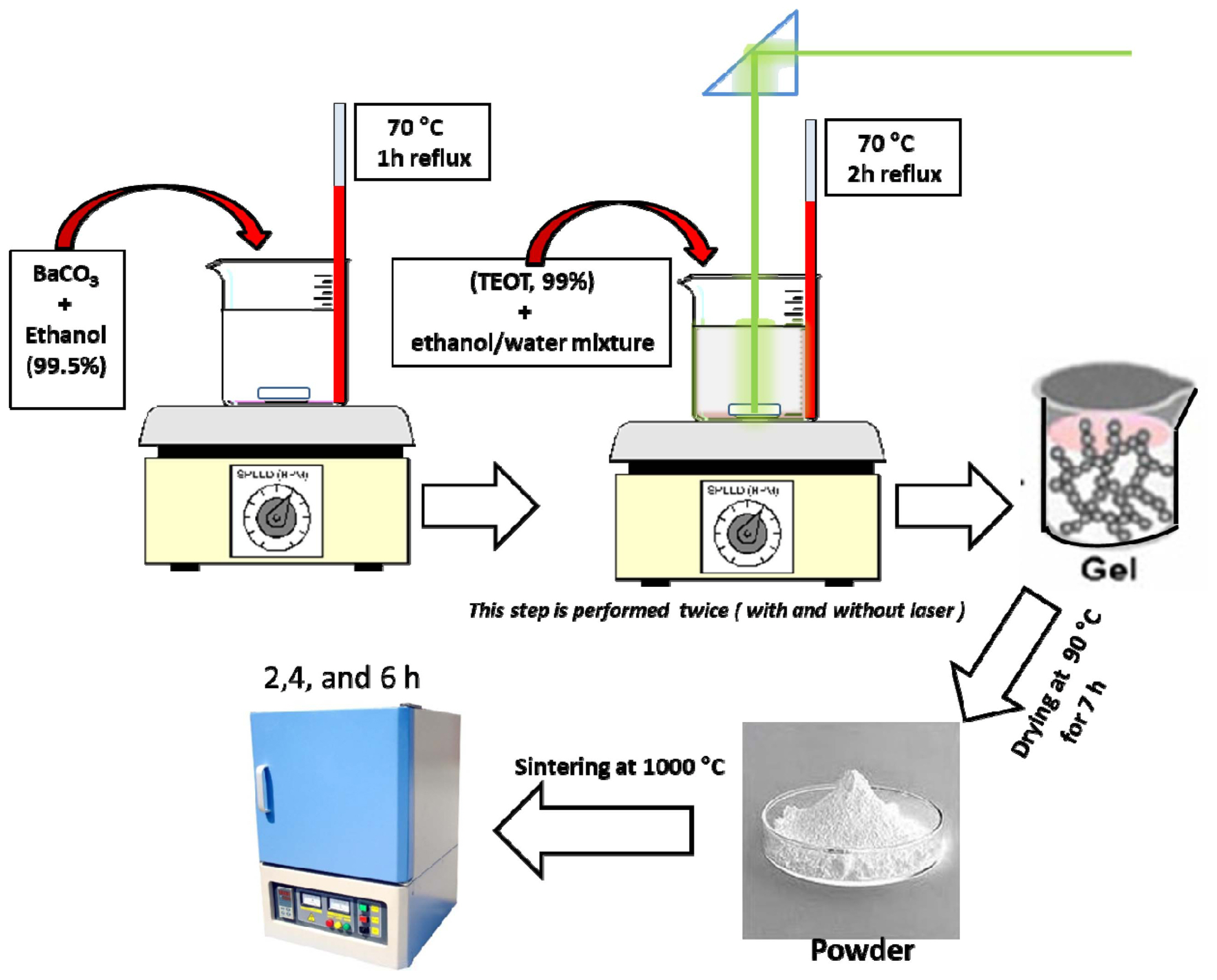
BT nano-particles were prepared by the sol-gel method. A complex alkoxide of barium-titanium was prepared as a precursor of the BT particles as follows: First, a solution (10−4 m3) that dissolved metallic barium (10 mmol) in ethanol was refluxed on magnetic starrer at 70°C for 1 h. Subsequently, TEOT (10 mmol) was added to the solution, which was refluxed for another 2 h—a transparent, complex alkoxide was obtained. To hydrolyze the complex alkoxide, the solution was mixed with an equal volume (10−4 m3) ethanol/water mixture. The material was then irradiated by green diode laser at 1000 mW power and 532 nm wavelength (GL1W), and—separately—red diode laser at 500 mW and 808 nm (RL0.5W). Each was fixed vertically for 2 h, as shown in Fig. 1. The BT nano-particles were prepared by calcining the BT precursor particle at 70°C (to remove the residual organics) and kept at 70°C for 10 h (Fig. 1). The solution turned opaque, indicating the formation of BT particles. By sintering at 1000°C for three durations (2, 4, and 6 h), BT nano-particles were deposited at different crystallite sizes. From the X-ray diffraction (XRD) patterns, crystallite size was calculated with the help of Scherer’s formula, t = 0.9λ/βcosθ; where λ = wavelength, β = full width at half maximum (FWHM), and θ = diffraction angle.22-25) The powder was pressed into discs of 7 mm diameter and 1 mm thickness at 20 MPa. The disks were then placed in a small silica crucible that was placed at the center of a larger crucible and covered with aluminum oxide.26)
2.3. Measurements
The BT particles were characterized by XRD. Particles were observed with a Zeiss LEO 912 OMEGA operated at 100 kV accelerating voltage. Crystal structures of the particles were measured with an X-ray diffractometer (Rigaku RU-200A) operated at 40 kV and 30 mA with CuKα radiation using a monochromator. Optical properties of the nanoparticles were studied by a UV-Visible spectrophotometer (UV2300II). The Fourier transmission infrared (FTIR) spectra of the samples were recorded by using FTIR (Shimadzu, model DF 803) in the wave range 400-4000 cm−1. A pellet of 7 mm diameter and 1 mm thickness was taken for the electrical properties study; thin, silver electrodes were screen printed onto opposite faces of the ceramic disk. For organic removal, printed disks were kept on an alumina plate and first fired at 500°C for 2 hours. To measure the capacitance of the sample it was then positioned between two copper electrodes that were connected to an automatic capacitance meter (RLC - meter model SRS).27)
3. Results and Discussion
3.1. Characteristics
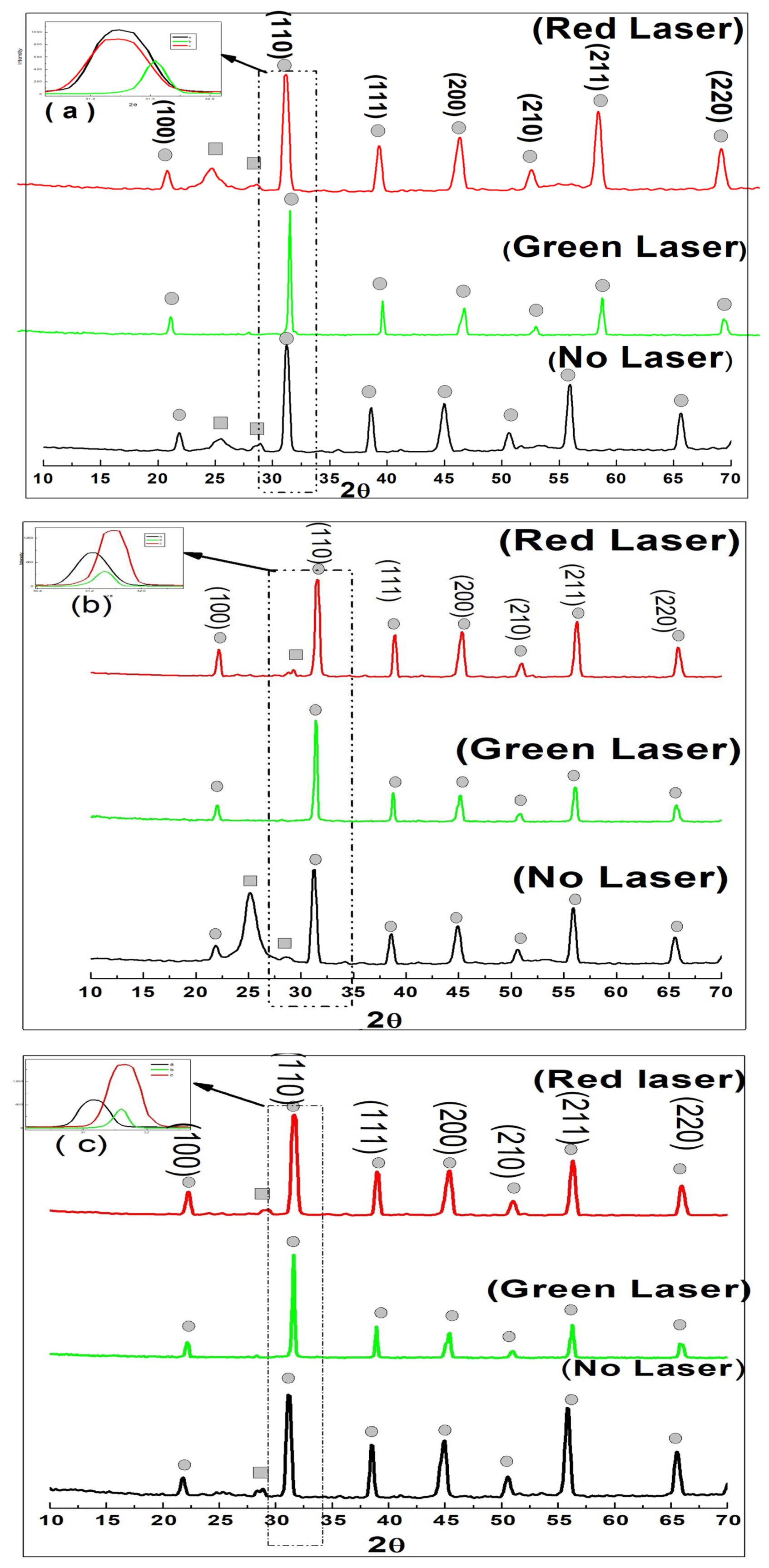
The BT particles were characterized by XRD analysis. Fig. 2 shows the XRD patterns of the BaTiO3 samples before irradiation (black line), after red laser irradiation (red line), and after green laser irradiation (green line). All samples were sintered at 1000°C—Figs. 2(a), 2(b), and 2(c) represent sintering times of 2, 4, and 6 h, respectively. The diffractogram indicates the formation of the tetragonal phase of BaTiO3, which is approved by the appearance of X-ray reflections at 2θ = 22.2, 31.4, 38.9, 45.39, 51.03, 56.3, and 65.95 (JCPDS 05-0626). The X-ray reflections at 2θ = 22.26, 31.69, 39.03, 45.09, and 56.35 are in correlation with the JCPDS standards (31-0174) for the sample and indicate the formation of cubic phase of BaTiO3. There is a peak shift observed at (110) toward higher angles as the sintering time increased for samples irradiated by green laser. It can be considered that the phase transformation lead to both an overall decrease in the lattice constants, as well as a contraction of the unit cells for these samples.28) Samples irradiated by the red laser shifted to higher values at 4 and 6 h sintering time. As shown in Fig. 2, peaks marked by circles represent pure BaTiO3, while rectangles represent BaCO3. It is remarkable that samples prepared under green laser irradiation presented no trace of BaCO3, for all sintering durations. For samples irradiated by red laser, as well as those without laser irradiation, increasing sintering time results in a decrease of BaCO3.
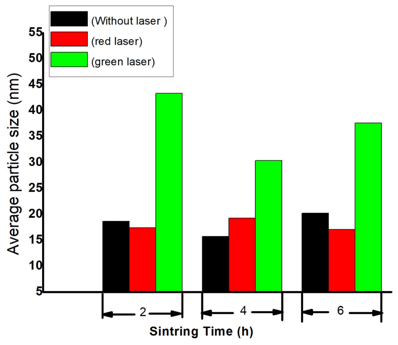
Average particle sizes—calculated by Scherer’s formula—are shown in Fig. 3, and illustrate that samples irradiated with green laser exhibit a higher grain size for all sintering durations.
3.2. Morphology
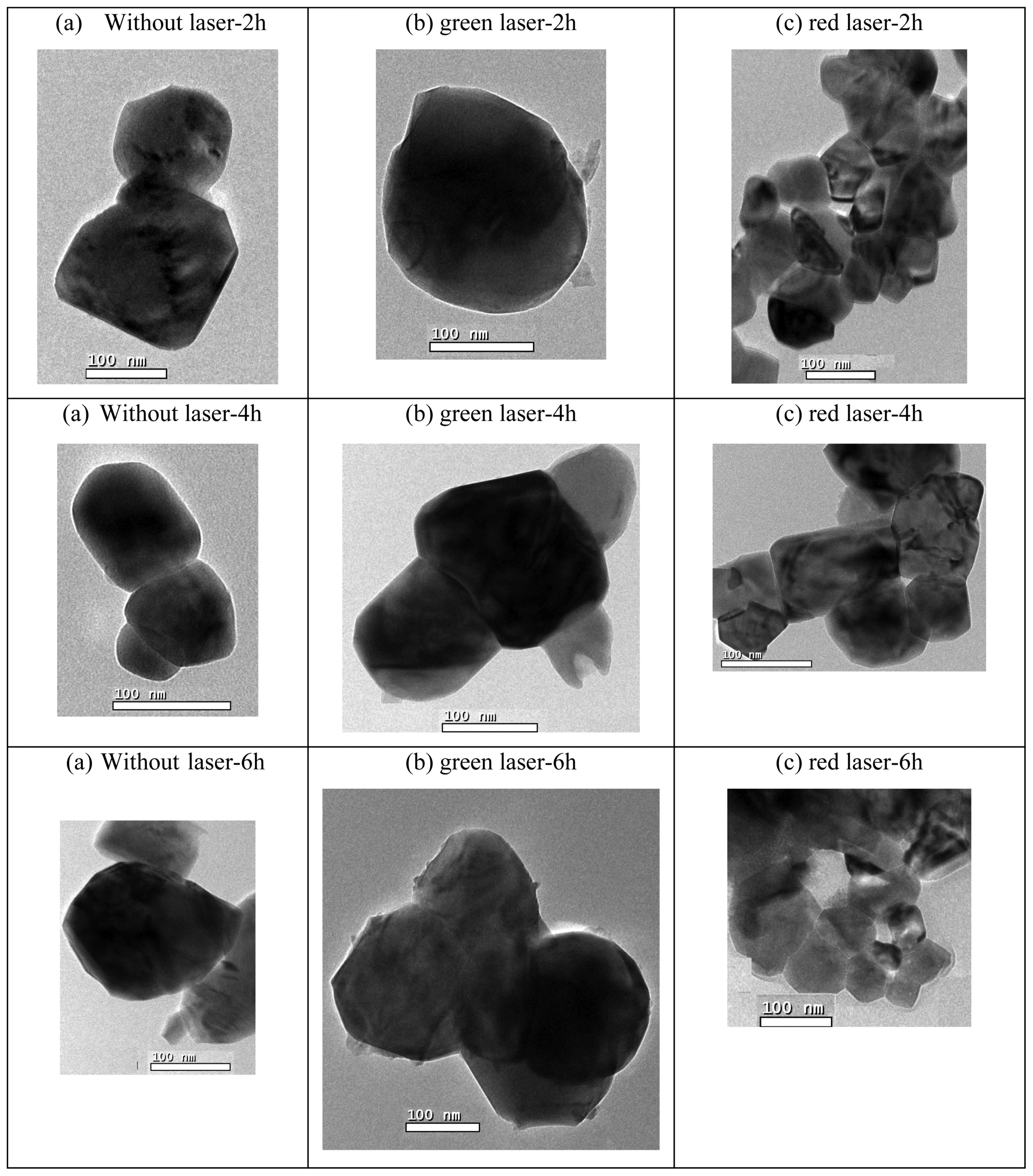
The structural morphology and size of the synthesized BT nanostructures were investigated using transmission electron microscopy (TEM). Fig. 4 shows the TEM images of the BT nanostructures sintered at 1000°C for 2, 4, and 6 h, before and after irradiation by green and red lasers. The images highlight that some particles agglomerated. It is also observed that particles prepared under green laser irradiation exhibit a higher average particle size for all sintering durations.
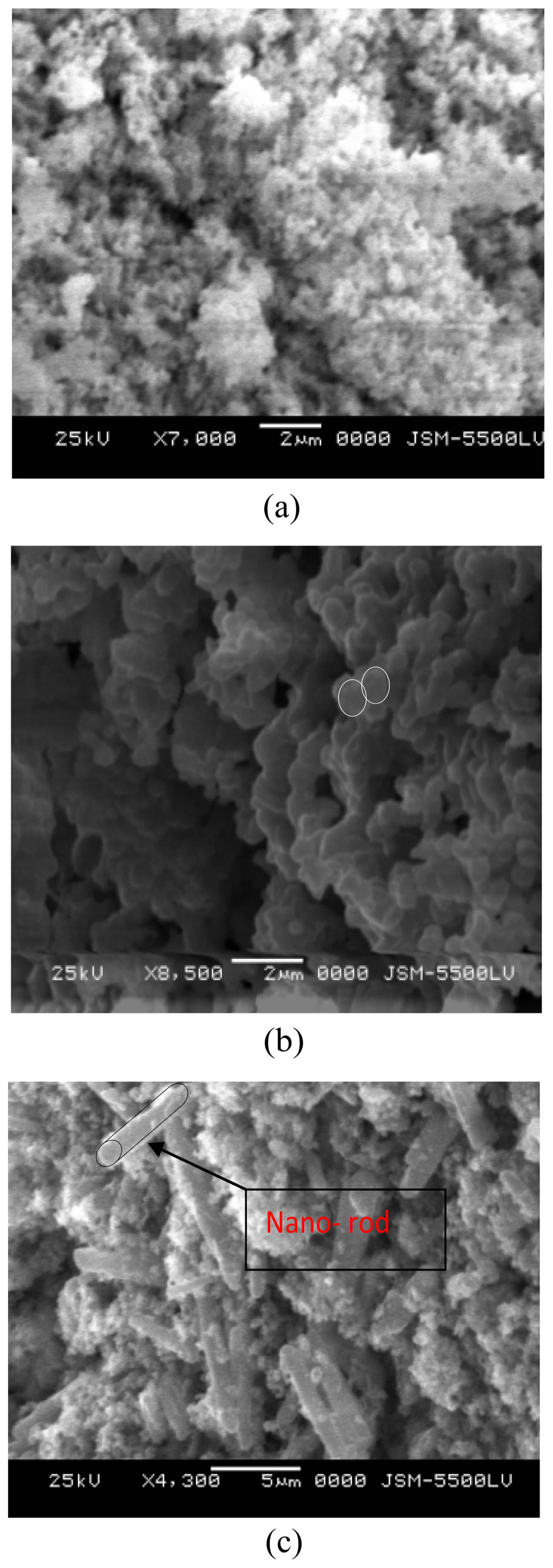
Figure 5 shows SEM (scanning electron microscope) images of BaTiO3 particles sintered at 1000°C for 2, 4, and 6 h, before and after green and red laser irradiation. Each laser appears to produce a different morphology. In the case of samples prepared under the red laser, some particles were transformed into a rod-like morphology (Fig. 5(c)), while samples prepared under green laser show spherical aggregates (Fig. 5(b)).
3.3. FTIR analysis
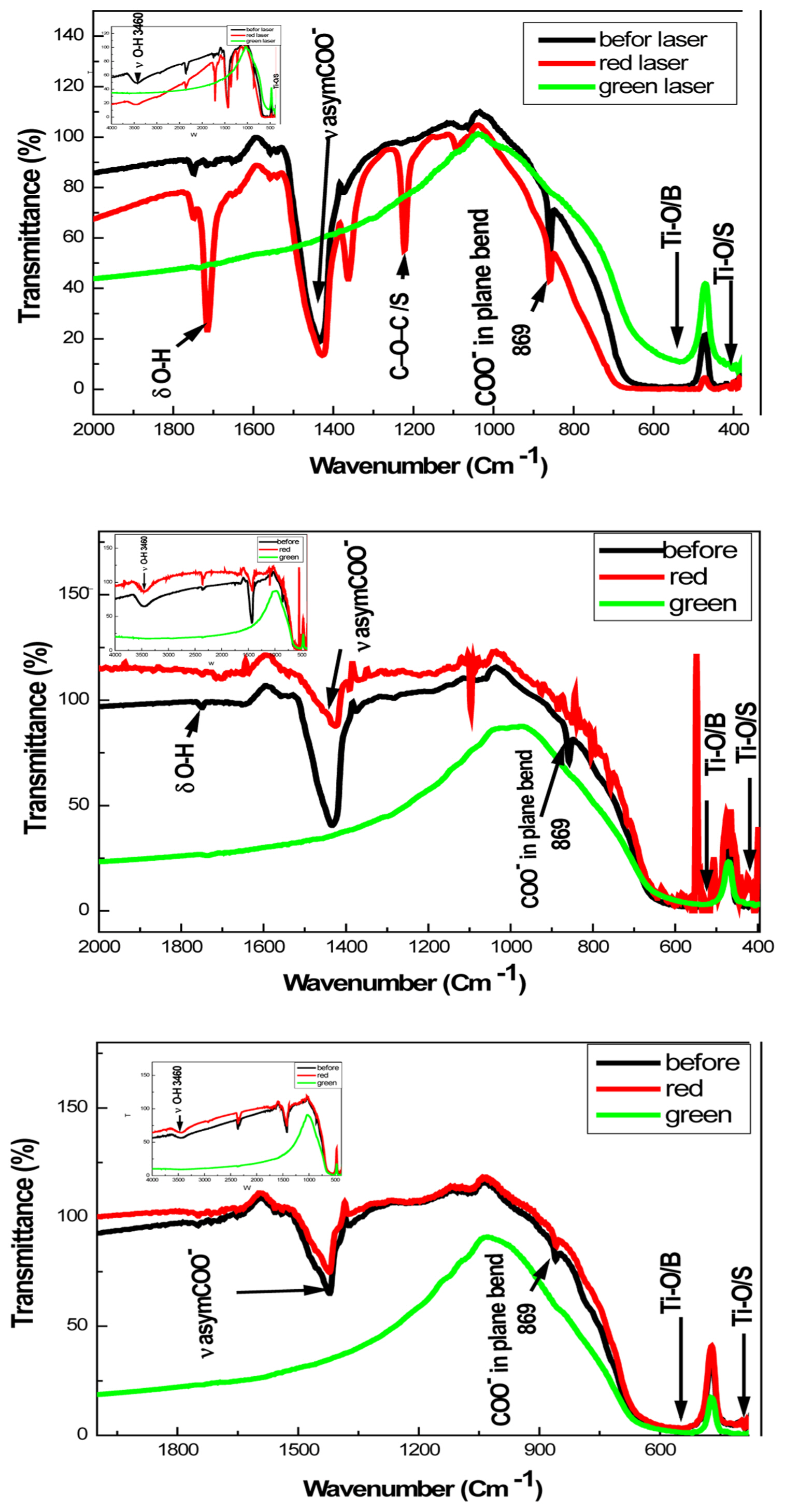
According to the literature, BaCO3 is the most common impurity of BaTiO3 powder prepared by any method. FTIR was found to be the most sensitive technique for BaCO3 detection. To check the purity of our obtained BaTiO3 powder, the FTIR spectra of the samples were noted and shown in Fig. 6. The FTIR spectra show a high purity for samples prepared under green laser irradiation, characterized by the presence of two broad bands centered at 540 cm−1, and below 500 cm−1—typical of the Ti-O stretching vibrations, and Ti-O bending vibrations (in BaTiO3), respectively.29) This result is compatible with that obtained by XRD.
The band at 869 cm−1 corresponds to the bending vibrations (in plane) characteristic of CO2. The peaks at 1240 cm−1 and 1051 cm−1 are attributed to C-O-C and C-O stretching patterns, respectively.30,31) The band at 1440 cm−1 can be interpreted as C-O vibration due to unavoidable traces of carbonate.32) The bands at 1630 cm−1 are due to OH deformation vibrations (δ). The bands at 3428 cm−1 (Fig. 6 inset) are due to OH stretching vibrations (ν).
3.4. Optical properties
The absorbance spectra of the obtained BaTiO3 at different sintering times are shown in Fig. 7.
In these spectra, there are minimal fluctuations in BaTiO3 samples prepared under green laser. This phenomenon may be the reason for its high transparency in the wavelength range from 300 nm to 600 nm.
3.5. Suggested interpretation
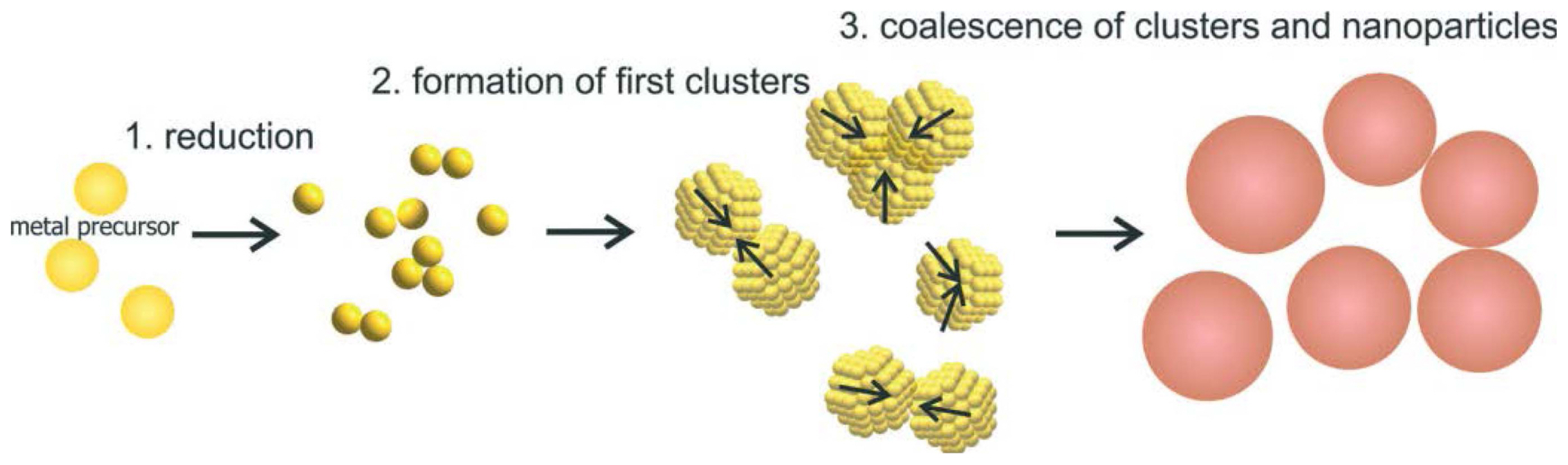
As described in the previous section, laser irradiation (particularly that of the green laser) has some effect on the structure, as well as the optical and electrical properties of BaTiO3. Molecules consist of atoms joined by chemical bonds. In large molecules (those with more than two atoms), the bonds may be weak, or strong, depending on the shape and atomic constituents of the molecule. The consequential general mechanism for nanoparticle growth with fast reducing agents can be described in three steps, as shown in Fig. 8. The first step is a fast reduction of the metal ions. Following the reduction, the second step is marked by the formation of the metal atoms into small clusters. In the third step, the clusters grow due to aggregation and coalescence until reaching a final size at which the particles have sufficiently stabilized.
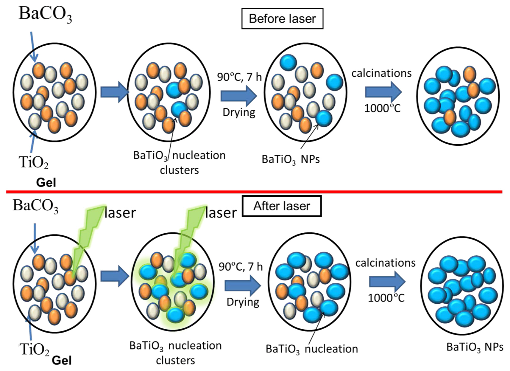
We suspect that the laser beam activates the primary nucleation of the BaTiO3 clusters, leading to more rapid formation of BaTiO3 molecules than without a laser. Based on this experimental result, the expected BaTiO3 formation process is concluded in Fig. 9—with and without laser irradiation—in four steps. The first step is gelation, the second step is to irradiate the gel clusters to activate the BaTiO3 molecules, the third step involves further nucleation of the BaTiO3 after drying to aerogel, and the last step leads to complete formation of BaTiO3 following calcination.
The photon energy produced by the 532 nm green diode laser is greater than that of the 808 nm red laser. This explains why the ideal, seeded BT growth process occurred under green laser irradiation, rather than the red laser.
3.6. Electrical behavior
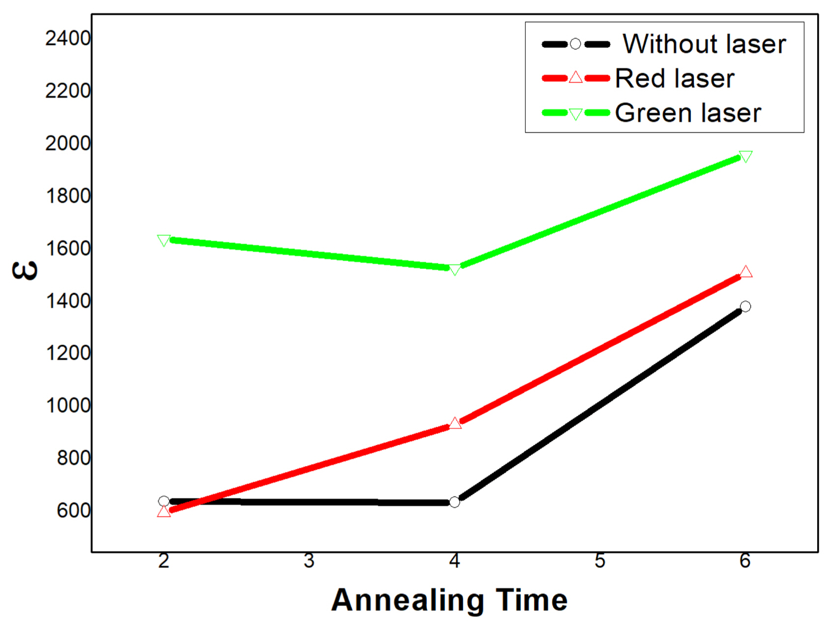
The dielectric constant was calculated using the standard equation for parallel plate capacitor, ɛr = C · d/(A · ɛo), where C is the measured capacitance, d is the sample thickness, A is the overlapping area of the capacitor plates, and ɛ∘ is the permittivity of the vacuum. Fig. 10 shows the temperature dependence of the dielectric constant for samples irradiated by red laser (red lines), green laser (green lines), and without laser irradiation (black lines). This measurement is performed for 2, 4, and 6 h sintering times. It can be observed that the relative permittivity is characterized by the higher dielectric constant values, as concluded in Fig. 11. It is also observed that the dielectric constant increases with sintering temperature. The observed maximum dielectric constant (ɛmax) and Curie temperature Tc of all studied samples are given in Table 1.
The increase in dielectric constant according to sintering temperature is known to be related to oxygen ions or oxygen vacancies created during the sintering process.34)
4. Conclusions
BaTiO3 samples were synthesized via sol-gel method and their structural, optical, and electrical properties were studied. XRD analysis shows the pure-phase formation in all samples of BT irradiated by a green laser diode (532 nm, 1000 mW) for different sintering durations at 1000°C. Low impurities of BaCO3 were observed for the other samples. TEM images shows that the laser beam application yields a larger grain size. TEM images show different morphologies for samples irradiated by laser. Expansion and deformation of the nano-dimensions are most apparent after green laser irradiation. Maximum electrical permittivity, ɛr, shows higher values for samples irradiated by the green laser than others. The maximum value (~ 1962) was observed at a sintering time of 6 h.